Human Anatomy Pelvis Muscles / Muscles Of The Abdomen Lower Back And Pelvis - It is also referred to as a ball and socket joint and is surrounded by muscles, ligaments, and tendons.
byAdmin-
0
Human Anatomy Pelvis Muscles / Muscles Of The Abdomen Lower Back And Pelvis - It is also referred to as a ball and socket joint and is surrounded by muscles, ligaments, and tendons.. Structures arteries of the pelvis and perineum bones of the pelvis and perineum fascia of the pelvis and perineum joints of the pelvis and perinium lymphatics of the pelvis and perineum muscles of the pelvis and perineum nerves of the pelvis and perineum topographical anatomy of pelvis and perineum veins… Of human anatomy and different types of motion, inspiring more realistic and energetic figurative art. Shop our carefully curated collection of high quality products today! Urinary, gi, reproductive system elements. This is a table of skeletal muscles of the human anatomy.
12 photos of the muscle anatomy pelvis. Some of the most important include the major digestive organs, the intestines. The pelvic floor muscles are the layer that supports the pelvic organs and spans the bottom of the pelvis. (2) the levator ani and the coccygeus, which together form the pelvic diaphragm and are associated with the pelvic viscera. The vertebral column of the lower back includes the five lumbar vertebrae, the sacrum, and the coccyx.
Pelvis And Perineum Anatomy Vessels Nerves Kenhub from thumbor.kenhub.com Pelvis anatomy collection at alibaba.com tailored to cater to the needs of medical students.pelvis anatomy products act as teaching aids for faculties at medical institutes.pelvis anatomy items are manufactured to simulate human anatomy and functioning accurately. The bones of the pelvis and lower back work together to support the body's weight, anchor the abdominal and hip muscles, and protect the delicate vital organs of the vertebral and abdominopelvic cavities. This muscle extends from the bony surfaces of the pelvis into the lesser sciatic foramen and also inserts on the greater trochanter. They have several functions, including helping to support the pelvic organs. See more ideas about anatomy, perineum, pelvis. The main focus of this article will be the pelvic floor muscles.on that topic, there are several important questions that need to be answered: Discover the muscle anatomy of every muscle group in the human body. It can be divided into the greater pelvis and the lesser pelvis.
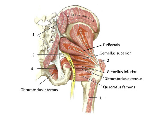
Pelvic diaphragm stretches from the pubic symphysis anteriorly, to the coccyx posteriorly, and is laterally attached to the medial surface of the obturator internus.
Learn vocabulary, terms, and more with flashcards, games, and other study tools. An important group of muscles in the pelvis is the pelvic floor. Males and females differ significantly in the anatomy of the pelvis: It can be described as one of the bodies diaphragms. Attached to the pelvis are muscles of the buttocks, the lower back, and the thighs. Terms in this set (54) bowl shaped, enclosed by true pelvis, continuous with abdominal cavity. Some of the most important include the major digestive organs, the intestines. The thigh bone or femur and the pelvis join to form the hip joint. The pelvis consists of the sacrum, the coccyx, the ischium, the ilium, and the pubis. Pelvic floor muscles located wholly within the pelvis The pelvis is a basin shaped bony structure formed by the combination of two pelvic bones (hip bones or innominate bones) and the sacrum. Pelvis anatomy collection at alibaba.com tailored to cater to the needs of medical students.pelvis anatomy products act as teaching aids for faculties at medical institutes.pelvis anatomy items are manufactured to simulate human anatomy and functioning accurately. This muscle extends from the bony surfaces of the pelvis into the lesser sciatic foramen and also inserts on the greater trochanter.
The intestines are supported by a series of muscles known as the pelvic. These muscles, including the gluteus maximus and the hamstrings, extend the thigh at the hip in support of the body's weight and propulsion. 12 photos of the muscle anatomy pelvis. The pelvic floor muscles provide foundational support for the intestines and bladder. An important group of muscles in the pelvis is the pelvic floor.
Functional Anatomy Of The Small Pelvic And Hip Muscles Completed Institute Of Basic Medical Sciences from www.med.uio.no Pelvis anatomy collection at alibaba.com tailored to cater to the needs of medical students.pelvis anatomy products act as teaching aids for faculties at medical institutes.pelvis anatomy items are manufactured to simulate human anatomy and functioning accurately. Cascade has served midwives and other healthcare professionals for over 40 years! Learn vocabulary, terms, and more with flashcards, games, and other study tools. The pelvic floor muscles are the layer that supports the pelvic organs and spans the bottom of the pelvis. To achieve both these tasks, the pelvic floor is composed of several overlapping sheets of muscles and connective tissues. The vertebral column of the lower back includes the five lumbar vertebrae, the sacrum, and the coccyx. Muscles that attach from the pelvis to the trunk and cross the lumbosacral joint muscles that attach from the pelvis to the thigh/leg and cross the hip joint pelvic floor muscles that are located wholly within the pelvis The pelvis is a basin shaped bony structure formed by the combination of two pelvic bones (hip bones or innominate bones) and the sacrum.
The small intestine is the longest part of the.
Learn vocabulary, terms, and more with flashcards, games, and other study tools. The intestines are supported by a series of muscles known as the pelvic. Structures arteries of the pelvis and perineum bones of the pelvis and perineum fascia of the pelvis and perineum joints of the pelvis and perinium lymphatics of the pelvis and perineum muscles of the pelvis and perineum nerves of the pelvis and perineum topographical anatomy of pelvis and perineum veins… Über 7 millionen englischsprachige bücher. The small intestine is the longest part of the. Inferior to pelvic cavity floor, boundaries form pelvic outlet. The pelvic floor is primarily made up of thick skeletal muscles along with nearby ligaments and their investing fascia. The pelvis is a basin shaped bony structure formed by the combination of two pelvic bones (hip bones or innominate bones) and the sacrum. If you're curious to know more, check out the full. It is strengthened and supported by several joints and ligaments. Cross sectional muscle anatomy of pelvis, ct pelvic muscle anatomy, mri pelvic muscle anatomy, pelvic muscle anatomy, pelvic muscular anatomy, human muscles, cross sectional muscle anatomy of pelvis, ct pelvic muscle anatomy, mri pelvic muscle anatomy, pelvic muscle anatomy, pelvic muscular anatomy. Shop our carefully curated collection of high quality products today! One is to close the pelvic and abdominal cavities and bear the load of the visceral organs;
The muscles within the pelvis may be divided into two groups: Discover the muscle anatomy of every muscle group in the human body. Pelvic floor muscles located wholly within the pelvis It can be described as one of the bodies diaphragms. The pelvic floor muscles include;
The Pelvic Floor Muscles Part 1 Basic Anatomy Youtube from i.ytimg.com The pelvis consists of the sacrum, the coccyx, the ischium, the ilium, and the pubis. The pelvic floor is primarily made up of thick skeletal muscles along with nearby ligaments and their investing fascia. This is a table of skeletal muscles of the human anatomy. Pelvic floor muscles located wholly within the pelvis The main focus of this article will be the pelvic floor muscles.on that topic, there are several important questions that need to be answered: Of human anatomy and different types of motion, inspiring more realistic and energetic figurative art. The pelvic region holds major organs under its layers of muscles. They are available as male, female or unisex manikins.
The pelvic floor has two inherently conflicting functions:
The pelvic region holds major organs under its layers of muscles. The femoral head is the distal (upper) end of the femur that inserts into the acetabulum of the hip joint. Terms in this set (54) bowl shaped, enclosed by true pelvis, continuous with abdominal cavity. (1) the obturator internus and the piriformis, which are muscles of the lower extremity, and will be described with these (pages 476 and 477); As it descends towards the knee, it is separated from the longest tubular bone, or shaft, by the femoral neck. See more ideas about anatomy, perineum, pelvis. The vertebral column of the lower back includes the five lumbar vertebrae, the sacrum, and the coccyx. Cross the ls joint onto the trunk 2. It is strengthened and supported by several joints and ligaments. Pelvis anatomy collection at alibaba.com tailored to cater to the needs of medical students.pelvis anatomy products act as teaching aids for faculties at medical institutes.pelvis anatomy items are manufactured to simulate human anatomy and functioning accurately. One is to close the pelvic and abdominal cavities and bear the load of the visceral organs; The muscles of the hip and thigh keep your hip joints strong and mighty, allowing for a wide range of hip movements. Attached to the pelvis are muscles of the buttocks, the lower back, and the thighs.
They are available as male, female or unisex manikins anatomy muscles pelvis. The thigh bone or femur and the pelvis join to form the hip joint.